胎儿超声心动图检查
胎儿超声心动图是怎么做的?
The test is typically performed by a specially trained ultrasound sonographer, and the images are interpreted by a 儿科心脏病专家 who specializes in fetal congenital heart disease. A limited evaluation of the fetal heart is possible during regular obstetric scanning and is appropriate for women at low risk. 然而, women who have one of the risk factors listed above should have a detailed fetal echocardiogram.
进行胎儿超声心动图检查有两种方法:
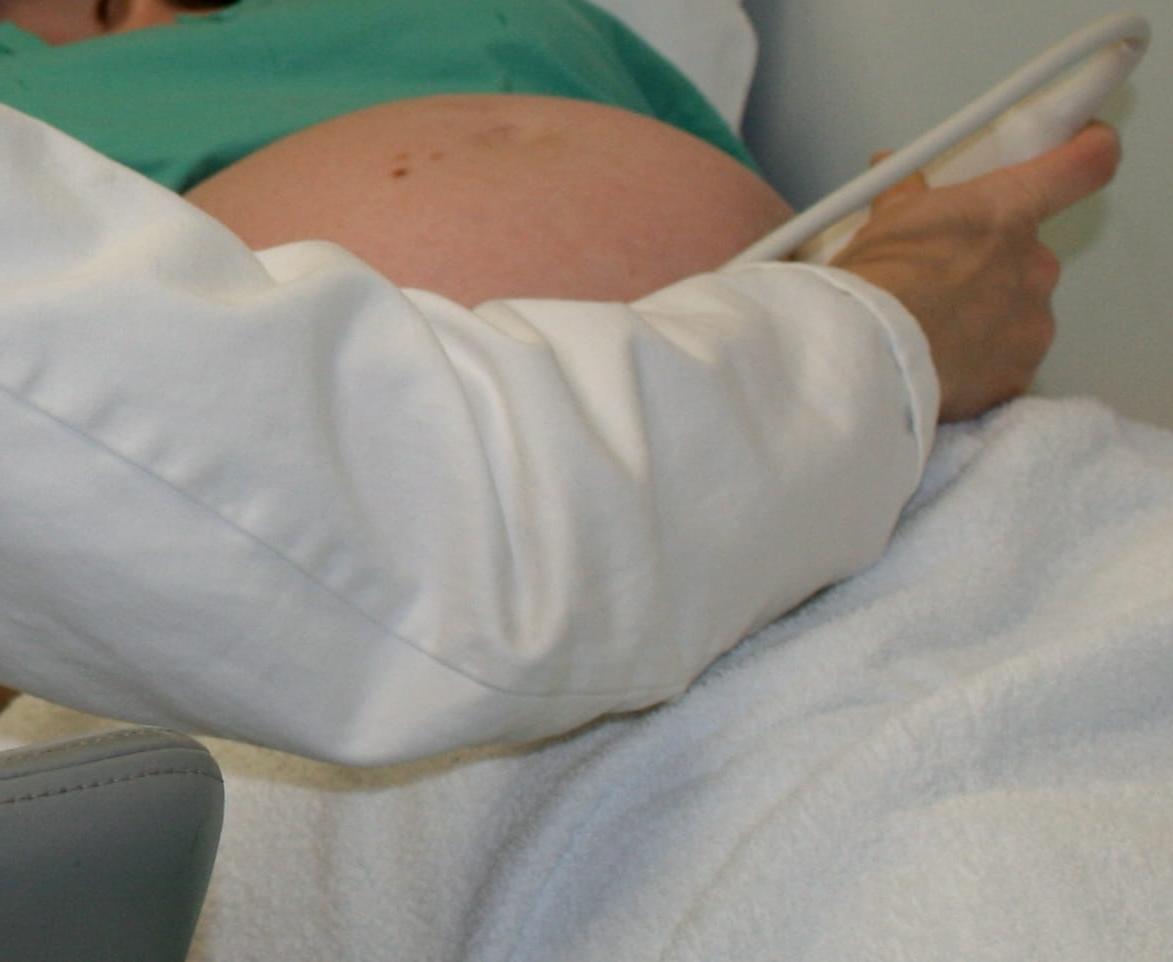
- 腹部超声: This is the most common form of ultrasound to evaluate the baby's heart. 一种凝胶被涂在妇女的腹部, the ultrasound probe is gently placed on the abdomen and pictures are taken. This test is not painful and causes no harm to the fetus. The test takes approximately 45–120 minutes depending on the complexity of the fetal heart.
- Endovaginal超声: 这种超声波通常在怀孕早期使用. A small ultrasound transducer is inserted into the vagina and rests against the back of the vagina. 然后可以拍摄胎儿心脏的照片.
考试需要做什么准备吗?
不像一些常规的产前超声波, a full bladder is not necessary for a fetal echocardiogram, 而且不需要特别的准备. You may wish to bring your medical information as that may be helpful to your health care team. The study can take anywhere from 30 minutes to over two hours.
测试什么时候完成?
The test is recommended at 18 to 22 weeks for all patients who have a 冠心病 以及冠心病患者的伴侣. The test may be completed earlier in pregnancy in parents with high rates of 冠心病 in their family or more severe cardiac defects.
结果会显示什么?
At your follow-up appointment your health care professional will discuss the results of your test. If the test is normal, no cardiac abnormalities were found. You will continue with routine pregnancy care and testing.
Abnormal results will tell you whether there is a problem with the way the fetus’s heart works or formed. It can also tell you whether there are abnormal heart rhythms (arrhythmias). 如果发现异常结果, you may need to undergo further testing and generally will be referred to a specialist who can treat the condition.
即使是在正常的研究中, you will be counseled that not all heart problems can be ruled out. 这是因为 胎儿血液循环 和出生后不同吗. 另外, very small holes between the lower chambers of the heart are hard to see and it may not be possible to see every part of the large blood vessels from the fetus’s heart.
The diagnosis of heart defect has significant implications for the overall health of the fetus. These health issues may have significant implications for the prognosis of the child and play a major role in helping you make decisions about your pregnancy. Your cardiologist will provide you with information about whether you need to worry about these other issues; however, when the concern is significant you will be referred to other members of the care team that work closely with the 儿科心脏病专家.
Other tests, procedures and referrals that may be needed include the following:
- 胎儿的超声波检查: A detailed ultrasound of the rest of the fetus is necessary to follow fetal growth, monitor fetal well-being and assess the rest of the fetus for abnormalities in other organs.
- 心脏磁共振成像: This test is becoming more widely available and can provide additional information about the overall health of the fetus and problems with certain organs.
- 羊膜穿刺术: This is a test performed to determine chromosomal and genetic disorders and certain birth defects. The test involves inserting a needle through the abdominal and uterine wall into the amniotic sac to retrieve a sample of amniotic fluid.
- 遗传咨询: Geneticists and genetic counselors provide an assessment of the likelihood of a genetic syndrome and possible abnormalities in other organs based on the heart diagnosis and other findings in the fetus. The geneticist can provide information to patients and their relatives concerning the consequences of a disorder, 发展或传播的可能性, 以及预防的方法, 治疗, 和管理.
- 专家咨询: Depending on the diagnoses you may be referred to other subspecialists including a maternal-fetal medicine specialist, 儿科心脏病专家, 新生儿学家和遗传学家.
- 社会工作者/护士: Someone who is familiar with heart disease in children can provide information about caring for a child with congenital heart disease.


